Newsletter

Newsletter : Vol. 3, Issue 2, 4 October 2022
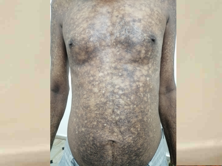
Vitamin B12 deficiency is common in India, as a majority of the population is vegetarian[1]. There are various cutaneous findings associated with cobalamin deficiency, the majority of which are more prevalent in patients with darker skin type. A retrospective and prospective study of 63 individuals with vitamin B12 deficiency related neurological syndromes in India showed that 41 % had skin and mucosal changes, with cheilitis in 31 %,hyperpigmentation in 19 %, hair changes in 9 %, angular cheilitis in 8 %, and vitiligo in 3 %[2]. We present a case of a 58 year old male who presented with an unusual pattern of cutaneous pigmentation on the whole body associated with vitamin B12 deficiency.
A 58 year old male came to us with complaints of shortness of breath on exertion, chest pain on exertion, easy fatigability, hyperpigmentation on face, reticulate hyperpigmentation on chest, upper and lower limbs of one month duration. on examination there was severe pallor with hyperpigmentation on face and reticulate pigmentation on chest, upper and lower limbs. Systemic examination was unremarkable. On laboratory examination, he had severe anaemia (hemoglobin 4.3 g/dL),leukopenia 2.6 × 10³/mm3 and thrombocytopenia (platelet count 50 × 103/mm3). His hematological indices—mean corpuscular volume (MCV) 100fL (70–86 fL), mean corpuscular hemoglobin 33.8 pg (25–35 pg), mean corpuscular hemoglobin concentration (MCHC) 33% (30%–36%)—suggested macrocytic anemia. 2Decho was normal .Vitamin B12 deficiency was suspected on clinical grounds and hematological data. His vitamin B12 levels were low (<50pg/ml)which was the cause for pancytopenia and reticulate hyperpigmentation. As he was non vegetarian, he was further evaluated for cause of vitamin B12 deficiency. Anti-parietal cell antibodies and anti-intrinsic factor antibody were sent. Anti-parietal cell antibodies were positive,-anti-intrinsic factor antibody was negative.so a diagnosis of vitamin B12 deficiency secondary to pernicious anemia was made. He was treated with vitamin B12 injections 1000mcg IM od for 7days,f/b 1000mcg IM once a week, f/b 1000mcg IM once a month, iron and folic acid supplements. His hematological indices normalized one month after treatment (hemoglobin:9.5 g/dL; MCV 90; MCH 33 pg; MCHC 33 %; TLC6100 /mm3, and platelet count 238× 103/mm3). The patient was continued on vit b12 supplementation once a month lifelong.
Cutaneous manifestations of vitamin B12 deficiency encompass a group of reversible but nonspecific findings. These include localized, generalized homogenous, reticulate, or honeycomb hyperpigmentation; angular stomatitis; glossitis, poliosis; and total melanonychia[3]. The cutaneous hyperpigmentation in our patient was present in a reticulate pattern along with typical hematological manifestations of vitamin B12 deficiency. Furthermore, the hematological manifestations improved after vitamin B12 supplementation. Such a pattern of pigmentation with vitamin B12 deficiency has not been described earlier.
Neuropsychiatric manifestations commonly associated with deficiency include myelopathy, neuropathy, dementia, neuropsychiatric abnormalities and rarely show optic nerve atrophy. SACD though a rare cause of myelopathy, is the most frequent clinical manifestation of B12 deficiency[4]. Our patient had no neurologic manifestations.
The generally accepted mechanism of this pigmentation is an increase in the melanin synthesis[5]. The other hypothesis proposed are 1) Deficiency of vitamin B12 decreases the level of reduced glutathione, which activate tyrosinase and thus leads to transfer to melanosomes. 2) Defect in the melanin transfer between melanocytes and keratinocytes, resulting in pigmentary incontinence[5].
Pernicious anemia (PA) is megaloblastic anemia that results from a deficiency in cobalamin (vitamin B12) due to a deficit of intrinsic factor (IF). Intrinsic factor is a glycoprotein that binds cobalamin and therefore enables its absorption at the terminal ileum. The disease is often described as an autoimmune disorder due to the findings of gastric autoantibodies directed against both IF and parietal cells. Pernicious anemia also correlates with other autoimmune diseases and as well as genetic diseases. Antibodies to intrinsic factor can be of two types. One is the blocking type against the B12 binding site present in 70% of patients, and the other is antibodies to parietal cells present in 90% of patients, but it is less specific[6]. Our patient had positive anti-parietal cell antibodies and negative anti intrinsic factor antibody so he was advised lifelong vitamin B12 supplementation.
REFERENCES
1.Antony AC. Prevalence of cobalamin (vitamin B-12) and folate deficiency in India: Audi alteram partem. Am J Clin Nutr. 2001;74:157–9.
2.Aaron S, Kumar S, Vijayan J, Jacob J, Alexander M, Gnanamuthu C. Clinical and laboratory features and response to treatment in
patients presenting with vitamin B12 deficiency-related neurological syndromes. Neurol India, 2005; 53(1):55-8
3.Kaur S, Goraya JS. Dermatologic findings of vitamin B12
deficiency in infants. Pediatr Dermatol. 2018;35(6):796-799.
4. Lee GR. Pernicious anemia and other causes of vitamin B12 (cobalamin) deficiency. In: Lee GR, Foerster J, Lukens J, Paraskevas F, Greer JP, Rodgers GM, editors. Wintrobe’s clinical hematology. 10. Baltimore: Lippincott Williams; 1999. pp. 941–964.
5.Mori K, Ando I, Kukita A. Generalized hyperpigmentation of the skin due to vitamin B12 deficiency. J Dermatol 2001; 28: 282-285.
6.LahnerE,AnnibaleB.Pernicious anemia: new insights from a gastroenterological point of view. World J Gastroenterol. 2009 Nov 07;15(41):5121-8.

| YOUNG DOCTORS MOVEMENT
| STATE LEADERS
| FMPC 2022
| AFPI
| The Spice Route India
| Editorial Board
| Executive
© All rights reserved 2022. Spice Route India 2022
Contact us at TheSpiceRouteIndia@gmail.com